【Nature】 Major Breakthrough in Asymmetric Protocell Division by Yan Qiao and Shu Wang’s Collaborative Team
Controlled division of artificial cells remains a key challenge in mimicking cellular behaviors. To date, researchers have induced vesicular protocell division by modulating membrane tension, membrane fluidity, and osmotic pressure through approaches such as protein machinery, external shear forces, light stimulation, ionic strength regulation, and thermal gradients. Symmetric division in emulsion and coacervate droplet systems has also been achieved through mechanisms including thermal gradients, dissipative self-assembly, wetting energy, and chemical reactions. In contrast, asymmetric cell division is a fundamental process in living systems that underpins cell differentiation, development, and functional diversification. However, owing to the lack of intricate spatiotemporal regulation and structural reorganization involved, reproducing asymmetric division in artificial cell systems has remained highly challenging.
With support from the National Natural Science Foundation of China, the Chinese Academy of Sciences, and the Beijing National Laboratory for Molecular Sciences, QIAO Yan’ group at the Laboratory of Polymer Physics and Chemistry has been dedicated to the construction of artificial cells and their cell-mimetic behaviors, achieving a series of significant advances (Nat. Chem. 2024, 16, 158–167; Nat. Chem. 2025, 17, 986–996; Nat. Commun. 2025, 16, 10554; J. Am. Chem. Soc. 2025, 147, 45004–45014).
Recently, the collaborative team led by Prof. QIAO Yan and WANG Shu, together with scientists from China and the U.K., published a research paper entitled “Asymmetric splitting in dividing lipid-nucleotide multilamellar droplets” in Nature. The research team developed an innovative strategy for asymmetric division in artificial cells based on transient chemical heterogeneity and interfacial energy gradients, establishing a structured droplet-based artificial cell model with autonomous asymmetric division capability (Figure). This system enabled the spontaneous division of a single parent droplet into two daughter protocells (an inner core droplet and an outer shell vesicle) with distinct morphologies and properties.
The research team constructed an artificial cell model based on lamellar liquid crystalline droplets formed by the self-assembly of lipid molecules and nucleotides. Upon catalysis by alkaline phosphatase, a localized caveola initially emerged on the droplet surface. As the enzymatic reaction progressed, the caveola propagated circumferentially around the droplet, accompanied by the emergence of a distinct shell–core interface within the structure. Once the caveola opening angle exceeded a critical threshold, the inner droplet core was extruded. Simultaneously, the detached shell underwent structural relaxation and edge closure, ultimately forming a multilamellar vesicular structure enclosing an internal aqueous lumen. As a result, a single parent droplet divided into two daughter protocells with markedly distinct structures, compositions, and properties.
The results demonstrated that the enzyme was predominantly enriched on the droplet surface, where the dephosphorylation reaction increased the interlamellar spacing of the droplets and induced structural instability within the droplets. Under non-enzymatic conditions, the introduction of multivalent ions or modulation of pH also triggered droplet division. These findings not only identified electrostatic shielding as the key driving force for asymmetric division but also demonstrated that the division mechanism was broadly applicable, which arises from the synergistic interplay between transient chemical inhomogeneity and interfacial energy gradients. Furthermore, the lamellar liquid crystalline structure and the minor structural defects within the layers were critical for droplet division, whereas structurally disordered droplets underwent only uniform disintegration.
Further studies revealed that the daughter droplets retained enzymatic activity and molecular transport capability, whereas the daughter vesicles gradually lost internal organization, released encapsulated molecules, and exhibit a decrease in pH due to ongoing dephosphorylation. These findings indicated that asymmetric division generated distinct chemical microenvironments within the progeny structures, thereby providing a basis for subsequent functional differentiation.
This research not only offers a new experimental model for understanding the emergence of life-like functions, but also lays an important foundation for constructing sophisticated artificial cell systems capable of autonomous proliferation, differentiation, and evolution.
These findings were recently published in Nature (Nature 2026. DOI: 10.1038/s41586-026-10489-5). The co-first authors are MENG He and JIA Liyan from the Institute of Chemistry, and the corresponding authors are Prof. QIAO Yan (Institute of Chemistry), Prof. WANG Shu (Institute of Chemistry), Prof. LIN Yiyang (Beijing University of Chemical Technology), and Prof. Stephen Mann (University of Bristol). Professor QIU Dong from the Institute of Chemistry provided key support for small-angle/wide-angle X-ray scattering experiments. This work also received technical support from the Key Laboratory of Engineering Plastics (SAXS/WAXS) and the Huairou Research Center (STED super-resolution microscopy system) at the Institute of Chemistry.
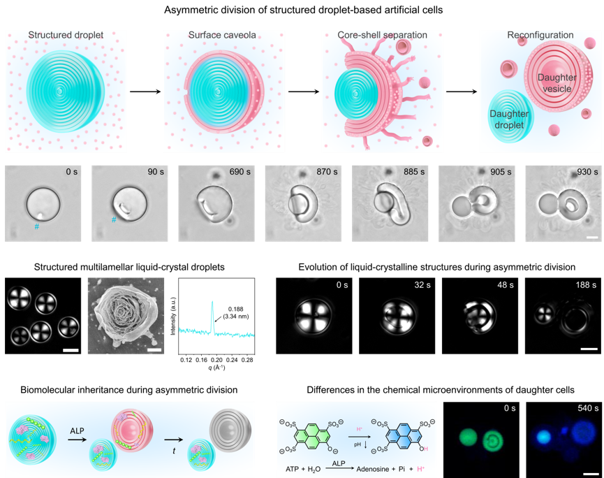
Figure. Asymmetric division of liquid crystalline droplet-based artificial cells. (Image by QIAO Yan)
Contact:
Prof. QIAO Yan
Institute of Chemistry, Chinese Academy of Sciences
Email: yanqiao@iccas.ac.cn