Researchers Develop Biomimetic Membranes for Coacervate Protocells
Coacervates formed by liquid-liquid phase separation have been investigated as protocells or synthetic cells due to their molecular crowded interior, guest molecular recruitment and dynamic adjustable assembly ability.
In a study published in Journal of the American Chemical Society, the research group led by Prof. QIAO Yan at the Institute of Chemistry of the Chinese Academy of Sciences, developed a plant cell-inspired membranization strategy to protect coacervate microdroplets with a layer of polysaccharide.
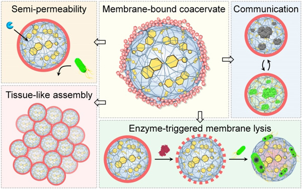
The membrane-bound coacervate protocells are presented as core-shell microcompartments (dextran membrane and coacervate droplet lumen), which are achieved by regulating charge-dipole interaction, hydrogen bonding, and steric hindrance effect. The membranized coacervate protocells show increased structural stability and controllable size evolution, allowing us to construct communicating protocell populations with membrane-gated permeability and enzyme-active bacterial predation. Moreover, the membrane-bound coacervates were further assembled into tissue-like superstructures, exhibiting efficient signal communication across microcompartments containing different enzymes.
This membrane-bound coacervate protocells provides a new platform for developing synthetic cell mimics with sophisticated life-like behaviour, and constructing hierarchical structures with multiple sub-microcompartments.

Biomimetic polysaccharide membrane for coacervate protocells (Image by Prof. QIAO Yan)
Contact:
Prof. QIAO Yan
Institute of Chemistry, Chinese Academy of Sciences
Email: yanqiao@iccas.ac.cn


