Fluorescent probes are powerful tools in the biological research and other fields due to their high sensitivity and specificity, fast response, and potential for real-time detection. The development of novel fluorescent probes has received more considerable attention in recent years.
Supported by the Ministry of Science and Technology of China, the National Natural Science Foundation of China and the Chinese Academy of Sciences, Professor Guoqiang Yang’s group in the Key Laboratory of Photochemistry of Chinese Academy of Sciences has studied on the synthesis of new fluorescent materials and the design of functional devices over the past years. A series of well-designed fluorescent molecules with high fluorescence quantum efficiency have been utilized as probes for fluoride ions and mercury ions in water (Angew. Chem. Int. Ed. 2010, 49 (29), 4915-4918, Anal. Chem. 2013, 85 (8), 4113-4119), temperature detection in different systems (Angew. Chem. Int. Ed. 2011, 50 (35), 8072-8076; Adv. Funct. Mater. 2013, 23 (3), 340-345; Chem. Comm. 2014, 50, 2778-2780.) and pH value in vivo (Chem. Comm. 2014,50, 8787—8790)
Recently, this group designed and synthesized a novel triarylboron derivative with hydrophilic, lipophilic and amphiphilic three-block structure, and utilized it as a fluorescent indicator for monitoring ATP levels in vitro and in vivo. The ATP-induced finite aggregation significantly enhances its fluorescence with the interaction between the probes and ATP molecules. The high specificity for ATP, low cytotoxicity, good permeability and dispersibility suggest it to be a good imaging agent for ATP distribution and alteration tracking. The determination of ATP level in NIH/3T3 fibroblast cells has been successfully performed with the indicator by the imagings of fluorescence microscopy and FLIM methods. The results have been published in Angewandte Chemie, International Edition (Angew. Chem. Int. Ed. 2014, 53 (30), 7809-7813).
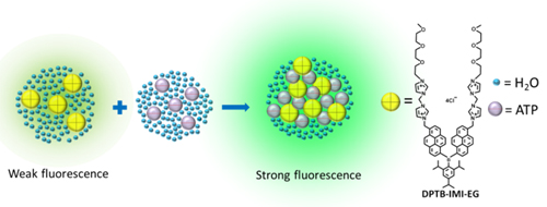
Figure 1 Schematic representation of the ATP indicator system

Figure 2 Confocal fluorescent images of live NIH/3T3 cells stained with DPTB-IMI-EG a) Fluorescence intensity image. b) Merged intensity and lifetime image